Toray develops CFRP part that improves diagnostic precision and reduces X-ray exposure
Toray has developed a radiolucent carbon fiber-reinforced plastic (CFRP) component that reduces X-ray radiation doses by 8%. This advancement enhances the use of CFRP in medical imaging applications, offering improved diagnostic precision with lower radiation exposure.
Toray Industries Inc. (Tokyo, Japan) has developed a radiolucent carbon fiber-reinforced plastic (CFRP) component designed to reduce X-ray radiation doses by 8% while maintaining or improving diagnostic image quality. This advancement is aimed at practical applications in X-ray examination equipment, such as mammography breast supports and cassettes.
X-ray examinations involve transmitting X-rays through the body to create images of internal structures, aiding early disease detection. However, these procedures carry risks of medical exposure, necessitating technologies that provide clear, low-noise diagnostic images without increasing radiation doses. In Japan, the Japan Network for Research and Information on Medical Exposure collects data on radiation doses and risk assessments, publishing diagnostic reference levels to optimize patient radiation exposure.
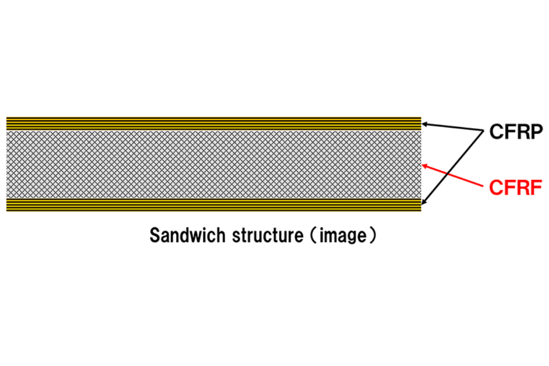
Protective CFRP components are used in X-ray equipment detectors due to their lightweight and radiolucent properties. Further improvement in radiolucency is crucial for obtaining diagnostic images without increasing radiation doses. A low-density sandwich structure with CFRP skins and a porous core is one effective approach. However, density variations in the porous core and component geometry can cause uneven transmitted X-ray intensity, potentially generating noise in diagnostic images. Thus, reducing radiation doses while enhancing diagnostic precision remains a challenge.
Toray has optimized a composite design for a sandwich structure using its carbon fiber-reinforced foam, achieving low density and high rigidity while minimizing density variations regardless of component geometry. Associate Professor Toru Negishi from Tokyo Metropolitan University evaluated these protective components under clinical conditions, confirming a 6% higher detective quantum efficiency compared to conventional CFRP. This enables obtaining diagnostic images of equal or greater clarity with an 8% reduction in X-ray dose. Implemented in examination equipment, such as mammography breast supports and cassettes, this component could enhance diagnostic precision and reduce medical exposure risks.
Source: Carbon Fiber Feed