Graphene oxide transforms stem cell spheroids into exosome factories for healing
Researchers in South Korea used graphene oxide to nearly double exosome production from induced pluripotent stem cells. This method enhanced angiogenesis, promoting faster wound healing in animal models.
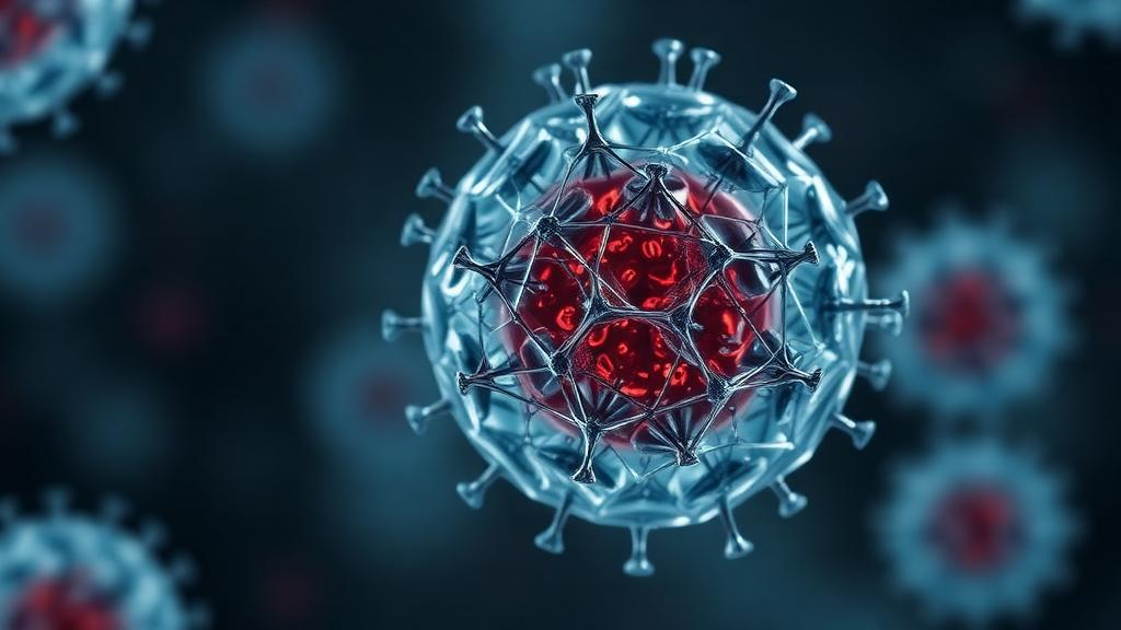
Graphene oxide has emerged as a pivotal material in enhancing the production of exosomes from induced pluripotent stem cells (iPSCs), according to recent research published in Materials Today Bio. Researchers in South Korea have demonstrated that incorporating graphene oxide into cell culture media leads to the formation of multiple small spheroids rather than a single large aggregate, nearly doubling exosome output. These exosomes have shown improved capabilities in promoting blood vessel growth, a key factor in tissue regeneration.
The study, led by Woochan Kim and Jangho Kim, involved detailed characterization of graphene oxide nanosheets, which measured approximately 300 to 400 nanometers across and 1.7 nanometers thick. The negatively charged, oxygen-rich surface of graphene oxide enhances its interaction with proteins and extracellular matrix molecules. When human iPSCs were cultured with graphene oxide, cell viability remained high, and no cytotoxicity was observed at concentrations up to 20 micrograms per milliliter.
Graphene oxide-treated cultures produced multiple smaller spheroids that persisted over five days, compared to conventional cultures that typically formed a single dominant spheroid per well. This structural change resulted in a significant increase in exosome production, with graphene oxide-engineered spheroids yielding 2.75 billion particles per million cells, compared to 1.4 billion from conventional spheroids. Importantly, the exosomes were free from graphene oxide contamination, as confirmed by Raman spectroscopy.
Functional assays revealed that exosomes from graphene oxide-engineered spheroids, termed GO-M-Exo, significantly enhanced angiogenic factor expression and improved outcomes in scratch wound and tube formation assays. The enhanced network formation was shown to be dependent on VEGFR2 signaling, as evidenced by the collapse of network formation upon the addition of a VEGFR2 inhibitor.
In vivo testing in a rat model of skin wounds demonstrated that GO-M-Exo led to nearly complete wound closure by day 14, outperforming untreated controls. Histological analysis showed improved granulation tissue, re-epithelialization, and microvessel density in the treated group, with no adverse inflammatory responses observed.
The researchers also extended their platform to produce lineage-specific exosomes by differentiating graphene oxide-engineered spheroids into vascular endothelial cells. These endothelial spheroids, called GO-MV, produced a higher yield of exosomes enriched in angiogenesis-related microRNAs and demonstrated superior performance in wound healing models.
The study highlights the potential of graphene oxide as a scalable and effective tool for enhancing exosome production in regenerative medicine. However, further research is needed to address long-term safety concerns and optimize purification methods to ensure clinical applicability.
Source: Graphene Feed