Researchers achieve high-res view of 2D materials with microscopic twist
Researchers at the University of Maryland developed infrared torsional force microscopy, enabling near-nanometer precision imaging of material surfaces responding to infrared light.
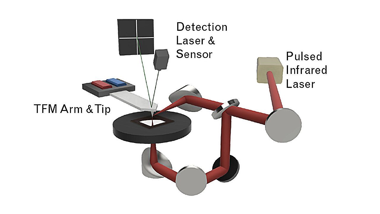
Researchers at the University of Maryland have developed a novel method to capture high-resolution images of two-dimensional materials, focusing on the intricate vibrations of carbon-based structures like graphene. This technique, known as infrared torsional force microscopy (TFM-IR), allows for near-nanometer precision in detecting both vertical and horizontal vibrations induced by infrared light. The study, published in Nature Communications, highlights how this method can reveal the unique vibrational fingerprints of materials, offering insights into their chemical composition and mechanical properties.
The TFM-IR technique builds on the principles of atomic force microscopy (AFM), which traditionally measures surface topography by dragging a tiny tip across a material. Unlike conventional AFM, which only detects vertical changes, TFM-IR employs a twisting motion of the AFM tip to capture horizontal variations. This advancement enables researchers to observe the subtle changes in materials like graphene, which are crucial for understanding their quantum properties.
In their experiments, the team used TFM-IR to study a double layer of graphene, a material known for its exceptional electrical and mechanical properties. By stacking two layers of graphene at a slight angle, they created a moiré material, which can exhibit superconductivity under specific conditions. The TFM-IR images revealed not only the lattice structure but also the distinct chemical bonds within and between the graphene layers, providing a comprehensive view of the material's response to infrared light.
The researchers also tested their method on mica, a mineral with well-known properties, to demonstrate the technique's ability to distinguish between vertical and horizontal vibrations. The results showed that TFM-IR could accurately map the vibrational patterns, aligning with theoretical predictions and simulations.
This new imaging capability is expected to be instrumental in the characterization and design of advanced materials, particularly in the semiconductor industry, where detecting defects at the nanoscale is critical. The TFM-IR technique offers a powerful tool for identifying the mechanical signatures of local chemistry, potentially guiding the development of next-generation nanoscale devices, including quantum sensors and photonic quantum computers.
Source: Graphene Feed