Tailored carbon nanofiber layer enhances ultra-thin fuel cell power density
Researchers found that a 25-micrometer carbon nanofiber film in ultra-thin fuel cells achieved 1.31 watts per square centimeter, surpassing both thicker and thinner film configurations.
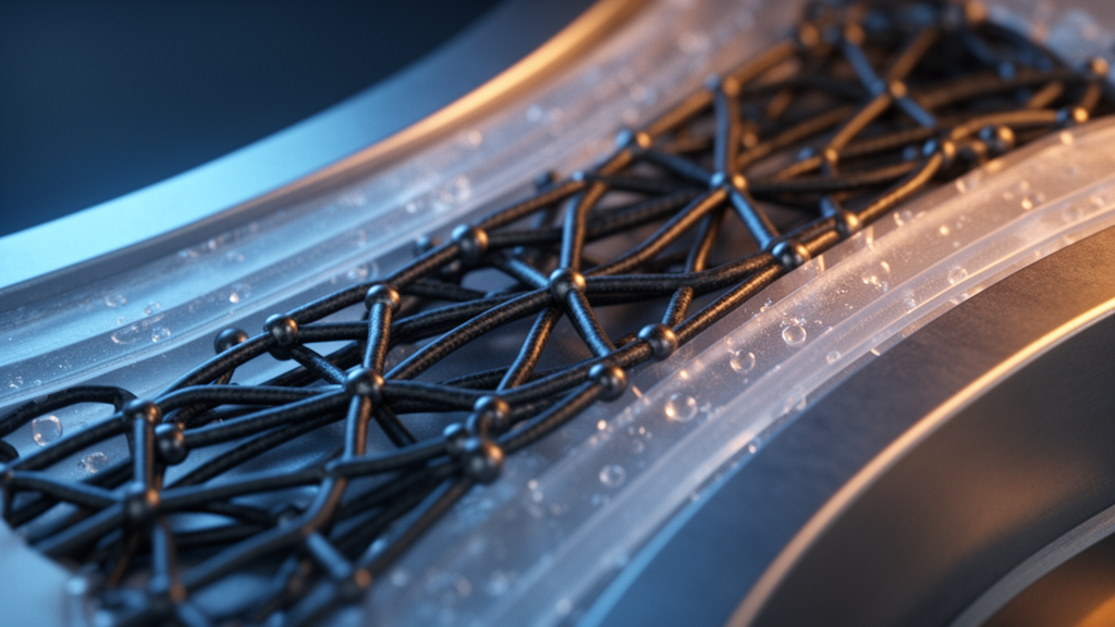
Researchers have conducted an experimental study on ultra-thin proton exchange membrane fuel cells, which eliminate traditional gas diffusion layers, by optimizing the thickness of an electrospun carbon nanofiber film to 25 micrometers. This configuration achieved a peak power density of 1.31 watts per square centimeter, marking a 21.3 percent improvement over a 34-micrometer film and outperforming thinner films of 15 and 8 micrometers, which reached 1.11 and 1.00 watts per square centimeter, respectively. The study highlights that removing conventional gas diffusion layers and using an open-pore metal foam flow field with a carbon nanofiber transition layer significantly reduces internal mass transport pathways and overall cell thickness. However, the effectiveness of this design hinges on maintaining a balance: the transition layer must be thin enough to minimize molecular diffusion resistance while being thick enough to protect the catalyst layer from the metal foam's localized contact pressures.
Proton exchange membrane fuel cells are crucial for clean energy conversion when paired with green hydrogen from renewable resources, offering high energy density and zero carbon emissions. Typically, the bipolar plate in these cells provides mechanical support, heat removal, electron conduction, and reactant gas distribution. Conventional channel-rib flow field designs within these plates create engineering challenges, such as mass transport resistance and manufacturing complexity. Thick gas diffusion layers and rib-channel structures lead to prolonged diffusion pathways and uneven reactant distribution, limiting volumetric power density. To address these issues, researchers have explored metal foam flow fields, which offer high porosity, low production costs, light weight, and excellent electrical and thermal conductivity. Previous studies confirmed that porous metal skeletons enhance fluid and thermal uniformity, but integrating metal foam directly with the membrane electrode assembly presents mechanical and transport challenges, necessitating a multifunctional buffer interface.
The research team implemented an electrospun carbon nanofiber film as a multifunctional transition interlayer between the membrane electrode assembly and the metal foam flow field to achieve an ultra-thin design without conventional gas diffusion layers. Previous work showed that removing the gas diffusion layer increased volumetric power density by 111 percent and reduced concentration losses at high current densities, but the impact of interlayer thickness was not fully understood. A carbon nanofiber film that is too thin shortens gas diffusion distances and reduces internal cell volume but risks structural integrity, leading to poor conformity and insufficient buffering against the metal skeleton. Conversely, a thicker nanofiber layer improves mechanical shielding and electrical contact but extends the reactant diffusion pathway, hindering oxygen delivery to active sites. The study aimed to clarify how varying the carbon nanofiber layer thickness affects mechanical protection, ohmic conductivity, reactant gas diffusion, and two-phase liquid water management during continuous cell operation.
The carbon nanofiber films were fabricated through precision electrospinning using polyacrylonitrile precursor solutions under controlled conditions, including spinning voltage, humidity, feed rate, and polymer concentration. By varying spinning times from 40 to 160 minutes, followed by stabilization at 240 degrees Celsius and graphitization at 1000 degrees Celsius, the researchers produced uniform carbon nanofiber membranes with thicknesses of 8, 15, 25, and 34 micrometers. To isolate the transition layer's effects, identical catalyst coated membranes were used in all test cells, employing a Gore M775.15 mechanically supported perfluorosulfonic acid membrane 15 micrometers thick. The anode and cathode catalyst layers used XC-72 carbon supports with platinum loadings of 0.05 and 0.40 milligrams per square centimeter, respectively, defining a 3-square-centimeter active area to emphasize through-plane transport contributions. Anode gas diffusion was provided by Toray 060 carbon paper, while graphite bipolar plates, gold-plated copper current collectors, and aluminum end plates secured with a 5 Newton-meter assembly bolt preload completed each single-cell assembly.
Electrochemical evaluations were conducted using a fuel cell testing bench in constant-current mode, with data acquisition increments of 0.05 amperes per square centimeter to generate triplicate polarization curves under ambient backpressure. High-purity hydrogen was supplied to the anode at 0.2 standard liters per minute, and compressed air was delivered to the cathode at 0.5 standard liters per minute, preheated and maintained at 70 degrees Celsius with 100 percent relative humidity via bubbling humidifiers. The polarization and ohmic-drop-free voltage curves showed a non-monotonic performance trajectory with decreasing nanofiber film thickness. Reducing film thickness from 34 to 25 micrometers increased maximum power density from 1.08 to 1.31 watts per square centimeter, driven by shorter oxygen transport paths. However, thinner membranes led to performance penalties: the 15-micrometer sample dropped by 15.3 percent to 1.11 watts per square centimeter, and the 8-micrometer membrane declined by 23.7 percent to 1.00 watts per square centimeter, indicating that extreme thinning undermines the electrochemical interface's functional integrity.
To identify the mechanisms behind performance losses in thinner layers, the authors conducted mathematical overpotential decomposition, separating total cell polarization into activation, ohmic, and concentration components using high-frequency resistance measurements. The ohmic overpotential did not decrease with reduced material thickness as expected; instead, it rose significantly in the thinnest films. This was attributed to two factors: thinner nanofiber sheets have lower lateral electrical conductivity, and their fragile structures suffer compression-induced fracturing against the metal foam struts, increasing contact resistance. Activation overpotentials also increased sharply as film thickness was reduced below 25 micrometers. The 25-micrometer film showed the lowest activation losses, while the 8-micrometer layer had the highest activation barrier across evaluated current ranges. This increase in activation overpotential indicated that very thin nanofiber interlayers fail to provide adequate mechanical buffering, allowing the metal foam to deform and damage the catalytic interface.
Confirmation of structural degradation was obtained through cyclic voltammetry measurements of the electrochemical active surface area and ex-situ scanning electron microscopy of post-operation catalyst layers. Electrochemical active surface area measurements showed a 30 percent reduction in catalyst active area in the cell with the 8-micrometer nanofiber film compared to the 25-micrometer configuration. Microscopic examination of the catalyst layer from the 8-micrometer cell revealed extensive surface gouging and indentations from contact with the nickel foam skeleton, with micrographs showing fragmented carbon nanofiber debris within structural craters. In contrast, the catalyst layer protected by the 34-micrometer film had fewer and milder surface impressions, proving that adequate nanofiber thickness cushions the membrane electrode assembly. When the nanofiber layer is too thin, skeletal intrusion crushes catalytic micropores, detaches ionomer networks, and destroys the three-phase boundary where electrons, protons, and reactant gases converge for the oxygen reduction reaction.
Detailed diagnostic testing using diluted oxygen mixtures of 1 percent dry molar oxygen in nitrogen across multiple chamber pressures enabled the separation of total oxygen transport resistance into pressure-dependent and pressure-independent regimes. At a chamber pressure of 150 kilopascals, total oxygen transport resistance initially fell from 0.595 seconds per meter for the 34-micrometer film to 0.531 seconds per meter for the 25-micrometer film, before increasing in thinner samples. Linear deconvolution showed that the pressure-dependent transport coefficient, reflecting Fickian molecular diffusion through porous voids, scaled with film thickness from 2.84 to 3.02, 3.14, and 3.28 milliseconds per meter per kilopascal across the 8, 15, 25, and 34-micrometer samples. Conversely, the pressure-independent transport resistance, encompassing Knudsen diffusion within catalyst micropores and solid-state dissolution through ionomer films, declined sharply from 0.206 seconds per meter at 8 micrometers to 0.102 at 15 micrometers and 0.060 at 25 micrometers. Since the mean free path of oxygen at cell operating conditions is about 6.46 nanometers—much smaller than the 350 to 1000 nanometer pores of the nanofiber film—Knudsen diffusion is negligible within the film itself. The increased transport resistance in thinner films is due to catalyst layer crushing that impedes nanoscale pore diffusion.
Electrochemical impedance spectroscopy from 100 millihertz to 100 kilohertz, combined with Distribution of Relaxation Times analysis, provided insights into dynamic operational limitations under varying current loads. Deconvoluted impedance spectra highlighted an overlapping activation and mass transport peak, designated as Peak P3, whose relaxation time and resistance followed a non-monotonic trajectory with operating current density. At low current densities of 0.4 amperes per square centimeter, charge transfer processes dominated, with the 8-micrometer sample producing the largest impedance arc due to its depleted electrochemically active surface area. As current increased to medium densities of 2.0 amperes per square centimeter, activation resistance diminished, but liquid water accumulation emerged. Contour plots of relaxation time distributions revealed critical turning points corresponding to pore flooding and effective diffusion suppression, which shifted systematically toward higher current densities.
Source: Biochar Feed