Coral-inspired scaffold reprograms immune cells to speed up bone regeneration
Researchers at the University of Pittsburgh have developed a scaffold using carbon nanotubes and nano-hydroxyapatite to enhance immune cell activity for faster bone regeneration.
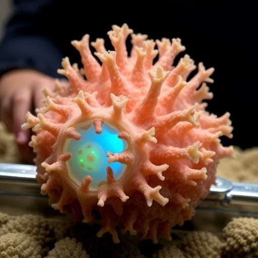
A coral-inspired scaffold integrating multi-walled carbon nanotubes (MWCNTs) with nano-hydroxyapatite (nHA) has demonstrated potential in enhancing bone regeneration for steroid-induced osteonecrosis of the femoral head (SONFH). This condition can lead to hip joint collapse. Researchers from Army Medical University and Sichuan University in China, in a study published in Bone Research, found that the 3D-printed biomaterial not only provided structural support but also modified the immune environment, promoting a reparative state in inflammatory macrophages. In rabbit models, this immune reprogramming correlated with increased blood vessel formation, enhanced bone-forming activity, and improved reconstruction of damaged femoral-head tissue.
SONFH is a severe complication of prolonged or high-dose glucocorticoid treatment, affecting 9% to 40% of patients. Glucocorticoids can disrupt blood supply, lipid metabolism, bone-cell activity, and immune regulation, weakening the femoral head. As the disease progresses, the femoral head may collapse, often necessitating hip replacement. Core decompression is commonly used in early stages but does not always restore the conditions needed for lasting repair.
The study identified persistent inflammation as a key barrier to bone healing post-surgery. Macrophages, immune cells involved in debris removal and tissue response coordination, can exist in different states. M1-like macrophages promote inflammation, while M2-like macrophages support tissue repair. In SONFH, macrophages remain in a pro-inflammatory state, inhibiting stem-cell function and new blood vessel growth, hindering bone formation.
To address this, the team designed a scaffold inspired by coral's pore network, using 3D printing to incorporate MWCNTs and nHA. MWCNTs, known for their mechanical, electrical, and surface properties, were used to influence macrophage behavior. nHA, resembling bone's mineral component, aimed to support cell attachment and differentiation into bone-forming cells.
The scaffold's porous architecture provided space for tissue infiltration and mimicked the 3D environment of native bone. Its composition influenced immune cells, encouraging macrophages to shift from an inflammatory to a reparative state, reducing inflammation and promoting mesenchymal stem cell activity. These cells are crucial for skeletal repair as they migrate to damaged areas and contribute to new bone formation.
The immunological effects were linked to activation of the PI3K-AKT signaling pathway, which influences cell behavior. The study associated this activation with macrophage reprogramming and the scaffold's regenerative response, suggesting it acts as a biological regulator rather than an inert replacement.
The scaffold also enhanced angiogenesis, crucial for repairing osteonecrotic bone by supplying oxygen, nutrients, and cells. Improved vascular development and reduced inflammation addressed recovery barriers, resulting in more extensive new bone formation in rabbit models compared to conventional treatments.
The study reflects a shift in regenerative medicine toward materials that actively communicate with living tissue. Successful bone repair may require controlling structural, vascular, and immunological processes. The coral-inspired scaffold targets inflammation as a central repair driver, offering a potential route to therapies that restore bone structure and its biological environment.
The research remains preclinical, with further studies needed to assess long-term safety, scaffold degradation, and the fate of MWCNTs. Consistent clinical-scale manufacturing and effectiveness in diverse medical conditions must also be evaluated. Despite these challenges, the study highlights the integration of immune reprogramming in bone engineering, emphasizing the need to address inflammation in regenerative therapies.
Source: Carbon Nanotubes Feed