Graphene reservoirs enable precise ice thickness control in cryo-EM
Researchers from Peking University, Tsinghua University, and the University of Hong Kong developed graphene reservoirs to precisely control ice thickness in cryo-electron microscopy, achieving uniform depths as thin as 15 nanometers.
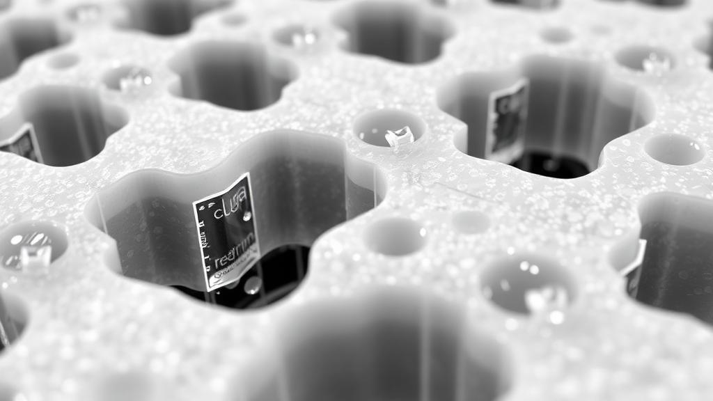
Researchers from Peking University, Tsinghua University, and the University of Hong Kong have developed a method utilizing graphene reservoirs to control ice thickness in cryo-electron microscopy (cryo-EM). This approach addresses a longstanding issue in structural biology by using sagging graphene to create nanometer-scale basins that precisely dictate the thickness of the vitreous ice layer. This layer, crucial for imaging, can now be controlled to prevent background noise and protein denaturation.
The study, published in Nature Methods, highlights that the instability of thin liquid films, due to van der Waals forces, has historically complicated ice thickness control. By exploiting the properties of graphene, the researchers engineered reservoirs that maintain a stable liquid film even during extended blotting times, enhancing the consistency of cryo-EM sample preparation.
The graphene reservoirs were fabricated using deep-ultraviolet lithography, achieving uniform sagging depths that correlate with hole diameters. This technique is compatible with existing grids like ANTcryo and Quantifoil, allowing for easy adoption in labs. The method demonstrated scalability, producing over 500 grids on a single wafer.
During blotting, the graphene reservoir grids maintained stable liquid films for up to 10 seconds, significantly longer than conventional methods. This stability resulted in uniform ice thickness, which is crucial for high-quality imaging. The technique also improved the performance of graphene sandwich specimens, providing more homogeneous ice thickness and reducing beam-induced motion.
The application of this method yielded significant improvements in structural biology results. For instance, the Ll.LtrB group II intron and SARS-CoV-2 spike protein achieved high-resolution reconstructions. The method also facilitated the imaging of small proteins, which are often drug-discovery targets, by providing uniform thin ice.
Beyond single-particle imaging, the researchers anticipate that graphene reservoir sandwiches will benefit other cryo-EM applications, such as time-resolved studies and in situ liquid electron microscopy. The method's compatibility with standard workflows and protocols suggests a low barrier to adoption, potentially transforming cryo-EM sample preparation.
Source: Graphene Feed