Pistachio shells converted into glowing carbon dots with antioxidant properties
Researchers used a five-minute microwave method to convert pistachio shell waste into fluorescent carbon dots with antioxidant, anticancer, and antibacterial properties. The process achieved an 85% yield.
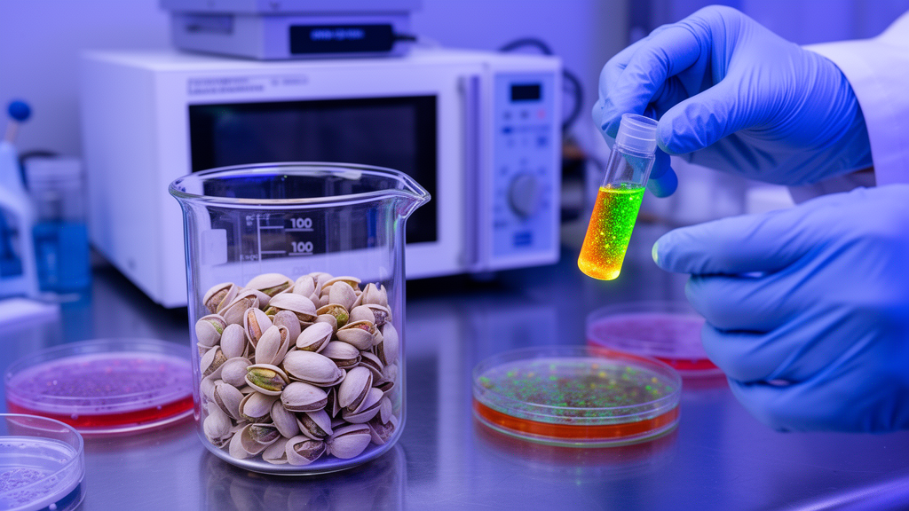
Researchers have developed a rapid method to convert pistachio shell waste into fluorescent carbon dots, showcasing potential applications in antioxidant, anticancer, and antibacterial domains. Utilizing a microwave-assisted process that takes just five minutes, the resulting nanomaterial exhibits strong radical-scavenging activity, compatibility with normal fibroblast cells, dose-dependent toxicity toward neuroblastoma cells, and the ability to inhibit the growth and biofilm formation of Escherichia coli and Staphylococcus aureus. This study, published as a full experimental report, highlights the use of agro-industrial waste as a renewable source for creating multifunctional carbon nanomaterials.
The synthesis process is straightforward. Pistachio shells from local agricultural waste were washed, dried at 70 degrees Celsius for twelve hours, and ground into powder. One gram of this powder was mixed with fifty milliliters of deionized water, stirred for thirty minutes, and then exposed to microwave irradiation at 300 watts and 90 degrees Celsius for five minutes. The carbon dots were purified through filtration, centrifugation, membrane passage, dialysis, and freeze-drying, achieving a mass yield of 85.0 plus or minus 0.8 percent. Three independent batches confirmed the reproducibility of the process, although the yield reflects purified product recovery rather than absolute carbon conversion due to unquantified volatile losses and water-soluble components.
The process's environmental impact was evaluated using the Eco-Scale approach, scoring 86.5, indicating an excellent green process at the laboratory scale. This score is attributed to the renewable waste-derived precursor, aqueous reaction medium, absence of additional chemicals, and short reaction time, with a microwave energy input of 90 kilojoules per batch. However, the authors caution that scaling up may present challenges such as changes in microwave penetration and reactor geometry, as well as the need to address purification steps like dialysis and freeze-drying.
Characterization of the carbon dots revealed their structural properties. High-resolution transmission electron microscopy showed quasi-spherical dots averaging five nanometers in diameter, with lattice fringes spaced at about 0.21 nanometers corresponding to the (100) plane of graphitic carbon. Nitrogen adsorption measurements indicated a specific surface area of 27 square meters per gram and a total pore volume of 0.08 cubic centimeters per gram, with mesoporous pores ranging from two to fifty nanometers. Fourier transform infrared spectroscopy and X-ray photoelectron spectroscopy confirmed an oxygen-rich surface with 62.44 percent carbon and 37.56 percent oxygen, featuring hydroxyl, carbonyl, and carboxyl functionalities that contribute to hydrophilicity and aqueous dispersibility.
Raman spectroscopy provided insights into the material's hybrid structure. The spectrum showed a D band at 1313 inverse centimeters, linked to disordered sp2 carbon, and a G band at 1597 inverse centimeters, associated with ordered graphitic domains. The intensity ratio of these bands was 1.0025, indicating a defect-rich framework with coexisting graphitic and amorphous regions. X-ray diffraction revealed a broad peak near 24 degrees, assigned to the (002) plane of graphitic carbon, with an interlayer spacing of 0.37 nanometers, larger than crystalline graphite, suggesting turbostratic layers and structural disorder due to oxygen functionalities.
Optically, the carbon dots emit blue fluorescence with a peak near 450 nanometers under 380 nanometer excitation, shifting to longer wavelengths with increasing excitation, indicating a distribution of emissive states. The fluorescence quantum yield was 17.2 percent, with an average fluorescence lifetime of 3.6 nanoseconds. The dots maintained 93 percent of their initial fluorescence after 360 minutes at pH 7.4 and 89 percent in serum-containing medium at 37 degrees Celsius, demonstrating stability under physiological conditions. Fluorescence varied across a pH range of two to twelve, suggesting ionizable surface groups contribute to emission, although contributions from the conjugated carbon core cannot be excluded.
Biological testing revealed a nuanced profile. In radical-scavenging assays, the dots achieved half-maximal inhibitory concentrations of 38.40 plus or minus 0.30 micrograms per milliliter against DPPH radicals and 35.10 plus or minus 0.90 micrograms per milliliter against ABTS radicals. At 100 micrograms per milliliter, they scavenged 92.2 plus or minus 1.7 percent of DPPH and 94.3 plus or minus 1.2 percent of ABTS radicals, performing moderately effectively compared to ascorbic acid and butylated hydroxytoluene. The activity is likely due to the oxygenated surface groups and partially graphitized framework, as pistachio biomass contains phenolic compounds and spectroscopic data cannot separate individual contributions.
In cell experiments, the dots were benign toward normal cells but toxic to cancer cells. L929 mouse fibroblasts exposed to concentrations up to 100 micrograms per milliliter for 24 hours retained 97 percent viability, maintaining their spindle-like morphology. In contrast, neuroblastoma Neuro-2a cells showed a concentration-dependent viability decline, from 97 percent at 5 micrograms per milliliter to 59 percent at 30 micrograms per milliliter, with the highest concentration significantly different from the control. A comparison with pyrrolidine dithiocarbamate confirmed substantial antiproliferative activity for the dots.
Mitochondrial and oxidative-stress measurements provided mechanistic insights. Pretreatment with the dots at 20 and 30 micrograms per milliliter reduced hydrogen-peroxide-induced reactive oxygen species in Neuro-2a cells, confirmed by fluorescence microscopy. Mitochondrial membrane potential was largely preserved at 20 micrograms per milliliter but dropped at 30 micrograms per milliliter, similar to the reference compound. These experiments demonstrate protection under oxidative stress and an association with mitochondrial perturbation at higher concentrations, though the precise mechanism of neuroblastoma cell death remains unidentified.
Antimicrobial testing completed the profile. E. coli growth was significantly suppressed at all concentrations down to 0.05 milligrams per milliliter, with complete inhibition at 0.1 milligrams per milliliter and above. Staphylococcus aureus required 0.4 milligrams per milliliter for complete inhibition, attributed to its thicker peptidoglycan layer. Biofilm formation was inhibited at 0.2 milligrams per milliliter for E. coli and 0.4 milligrams per milliliter for S. aureus. The metal-free nature of the dots avoids concerns of metal-ion release, and oxidative stress and cell-envelope interactions are proposed as mechanisms for the antibacterial effect, pending further studies. The study underscores the potential of pistachio shell waste as a sustainable source for multifunctional carbon nanomaterials and calls for further research to establish biological efficacy and safety.
Source: Emerging Nano Carbons